Application of simulated collagen fibrils in tissue engineering scaffolds
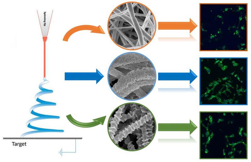
The ability to topographically mimic the surface features of collagen fibrils is an important step in the preparation of tissue engineering scaffolds. It is important to know which kinds of surface topographies of electrospun fibers are more favorable for cell growth. In this study, fibers with three kinds of hierarchical-structured surfaces were fabricated by electrospinning to mimic collagen fibrils. By combining thermally induced phase separation (TIPS) and non-solvent induced phase separation (NIPS), polycaprolactone (PCL) fibers with a porous surface were electrospun from PCL in a chloroform (CF)/dimethyl sulfoxide (DMSO) mixed solution. In addition, two additional types of fibrous membranes, with PCL fibers being the shish and decorated by PCL kebabs on the surface, were created by two different controlled homoepitaxic crystallization methods-the solution incubation method and the solvent evaporation method. It was found that the solvent evaporation method was more effective in forming kebabs and the primary optimal processing parameters were identified. The presence of pores on the fiber surfaces contributed to a much larger surface area and a higher total volume of pores. To investigate the cellular response on such scaffolds, 3T3 fibroblast cell and human umbilical vein endothelial cell (HUVEC) assays were conducted and the results indicated that both of the nanotopographies on the surfaces of the scaffolds improved cell viability and proliferation. Furthermore, the porous surface was more beneficial for enhancing cellular responses, which suggests better biocompatibility and greater potential to mimic collagen fibrils for tissue engineering application, and especially as scaffolds for endothelial layers in blood vessels. (c) 2017 Elsevier B.V. All rights reserved.